Back جسم بلعمي Arabic Fagosom BS Fagosoma Catalan Potravní vakuola Czech Phagosom German Fagosoma Spanish Fagosoma Basque Phagosome French Fagosoma Galician Fagosoma Italian

In cell biology, a phagosome is a vesicle formed around a particle engulfed by a phagocyte via phagocytosis. Professional phagocytes include macrophages, neutrophils, and dendritic cells (DCs).[1]
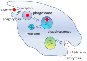
A phagosome is formed by the fusion of the cell membrane around a microorganism, a senescent cell or an apoptotic cell. Phagosomes have membrane-bound proteins to recruit and fuse with lysosomes to form mature phagolysosomes. The lysosomes contain hydrolytic enzymes and reactive oxygen species (ROS) which kill and digest the pathogens. Phagosomes can also form in non-professional phagocytes, but they can only engulf a smaller range of particles, and do not contain ROS. The useful materials (e.g. amino acids) from the digested particles are moved into the cytosol, and waste is removed by exocytosis. Phagosome formation is crucial for tissue homeostasis and both innate and adaptive host defense against pathogens.
However, some bacteria can exploit phagocytosis as an invasion strategy. They either reproduce inside of the phagolysosome (e.g. Coxiella spp.)[2] or escape into the cytoplasm before the phagosome fuses with the lysosome (e.g. Rickettsia spp.).[3] Many Mycobacteria, including Mycobacterium tuberculosis[4][5] and Mycobacterium avium paratuberculosis,[6] can manipulate the host macrophage to prevent lysosomes from fusing with phagosomes and creating mature phagolysosomes. Such incomplete maturation of the phagosome maintains an environment favorable to the pathogens inside it.[7]
- ^ Robinson & Babcock 1998, p. 187 and Ernst & Stendahl 2006, pp. 7–10
- ^ Hackstadt T, Williams JC (May 1981). "Biochemical stratagem for obligate parasitism of eukaryotic cells by Coxiella burnetii". Proceedings of the National Academy of Sciences of the United States of America. 78 (5): 3240–4. doi:10.1073/pnas.78.5.3240. PMC 319537. PMID 6942430.
- ^ Winkler HH (1990). "Rickettsia Species (As Organisms)". Annual Review of Microbiology. 44: 131–153. doi:10.1146/annurev.micro.44.1.131. PMID 2252380.
- ^ MacMicking JD, Taylor GA, McKinney JD (October 2003). "Immune control of tuberculosis by IFN-gamma-inducible LRG-47". Science. 302 (5645): 654–9. Bibcode:2003Sci...302..654M. doi:10.1126/science.1088063. PMID 14576437. S2CID 83944695.
- ^ Vandal OH, Pierini LM, Schnappinger D, Nathan CF, Ehrt S (August 2008). "A membrane protein preserves intrabacterial pH in intraphagosomal Mycobacterium tuberculosis". Nature Medicine. 14 (8): 849–54. doi:10.1038/nm.1795. PMC 2538620. PMID 18641659.
- ^ Kuehnel MP, Goethe R, Habermann A, Mueller E, Rohde M, Griffiths G, Valentin-Weigand P (August 2001). "Characterization of the intracellular survival of Mycobacterium avium ssp. paratuberculosis: phagosomal pH and fusogenicity in J774 macrophages compared with other mycobacteria". Cellular Microbiology. 3 (8): 551–66. doi:10.1046/j.1462-5822.2001.00139.x. PMID 11488816. S2CID 8962102.
- ^ Tessema MZ, Koets AP, Rutten VP, Gruys E (November 2001). "How does Mycobacterium avium subsp. paratuberculosis resist intracellular degradation?". The Veterinary Quarterly. 23 (4): 153–62. doi:10.1080/01652176.2001.9695105. PMID 11765232.