| Emesvirus zinderi | |
|---|---|

| |
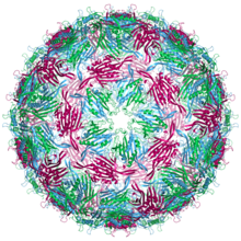
| Bacteriophage MS2 capsid structure. The three quasi-equivalent conformers are labelled blue (chain a), green (chain b) and magenta (chain c) | |
| Virus classification | |
| (unranked): | Virus |
| Realm: | Riboviria |
| Kingdom: | Orthornavirae |
| Phylum: | Lenarviricota |
| Class: | Leviviricetes |
| Order: | Norzivirales |
| Family: | Fiersviridae |
| Genus: | Emesvirus |
| Species: | Emesvirus zinderi
|
Bacteriophage MS2 (Emesvirus zinderi), commonly called MS2, is an icosahedral, positive-sense single-stranded RNA virus that infects the bacterium Escherichia coli and other members of the Enterobacteriaceae.[1] MS2 is a member of a family of closely related bacterial viruses that includes bacteriophage f2, bacteriophage Qβ, R17, and GA.[2]
It is small and contains a maturation protein, coat protein, and genomic RNA. It also has one of the smallest known genomes, encoding four proteins.
The MS2 lifecycle involves infecting bacteria with the fertility factor, enabling the virus to attach to the pilus, though the mechanism by which the virus's RNA enters the bacterium remains unknown. Once inside, the viral RNA starts functioning as a messenger RNA to produce viral proteins. MS2 replicates its plus-strand genome by creating a minus strand RNA as a template. The virus then assembles, and the bacterial cell lyses, releasing new viruses.
The virus was isolated in 1961 and its genome was the first to be fully sequenced, in 1976, providing a crucial understanding of genetic codes. In practical applications, MS2's structural components have been used to detect RNA in living cells. The virus is also under research for potential uses in drug delivery, tumor imaging, and light harvesting. Furthermore, because of its structural similarities to noroviruses, its preferred proliferation conditions, and its lack of pathogenicity to humans, MS2 serves as a substitute in studies of norovirus disease transmission.
- ^ van Duin J, Tsareva N (2006). "Single-stranded RNA phages. Chapter 15". In Calendar RL (ed.). The Bacteriophages (Second ed.). Oxford University Press. pp. 175–196. ISBN 978-0195148503.
- ^ Ni CZ, White CA, Mitchell RS, Wickersham J, Kodandapani R, Peabody DS, Ely KR (December 1996). "Crystal structure of the coat protein from the GA bacteriophage: model of the unassembled dimer". Protein Science. 5 (12): 2485–93. doi:10.1002/pro.5560051211. PMC 2143325. PMID 8976557.
