| Lobes of liver | |
|---|---|
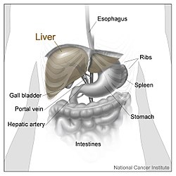
 The liver is divided into four lobes. This image shows the large right lobe and a smaller left lobe separated by the falciform ligament. | |
 1: Right lobe of liver 2: Left lobe of liver 3: Quadrate lobe of liver 4: Round ligament of liver 5: Falciform ligament 6: Caudate lobe of liver 7: Inferior vena cava 8: Common bile duct 9: Hepatic artery 10: Portal vein 11: Cystic duct 12: Common hepatic duct 13: Gallbladder | |
| Details | |
| Identifiers | |
| Latin | lobus hepatis |
| Anatomical terminology | |
In human anatomy, the liver is divided grossly into four parts or lobes: the right lobe, the left lobe, the caudate lobe, and the quadrate lobe. Seen from the front – the diaphragmatic surface – the liver is divided into two lobes: the right lobe and the left lobe. Viewed from the underside – the visceral surface – the other two smaller lobes, the caudate lobe and the quadrate lobe, are also visible.[1] The two smaller lobes, the caudate lobe and the quadrate lobe, are known as superficial or accessory lobes, and both are located on the underside of the right lobe.[2]
The falciform ligament, visible on the front of the liver, makes a superficial division of the right and left lobes of the liver. From the underside, the two additional lobes are located on the right lobe.[2] A line can be imagined running from the left of the vena cava and all the way forward to divide the liver and gallbladder into two halves.[3] This line is called Cantlie's line and is used to mark the division between the two lobes.[4]
Other anatomical landmarks exist, such as the ligamentum venosum and the round ligament of the liver (ligamentum teres), which further divide the left side of the liver in two sections. An important anatomical landmark, the porta hepatis, also known as the transverse fissure of the liver, divides this left portion into four segments, which can be numbered in Roman numerals starting at the caudate lobe as I in an anticlockwise manner. From this parietal view, seven segments can be seen, because the eighth segment is only visible in the visceral view.[5]

- ^ Karanjia N. "Anatomy of the Liver". Liver.co.uk. Retrieved 2015-06-26.
- ^ a b Moore KL, Dalley AF, Agur AM (2018). Clinically Oriented Anatomy (Eighth ed.). Philadelphia Baltimore New York London Buenos Aires Hong Kong Sydney Tokyo: Wolters Kluwer. pp. 493–498. ISBN 978-1-4963-5404-4.
- ^ Renz JF, Kinkhabwala M (2014). "Surgical Anatomy of the Liver". In Busuttil RW, Klintmalm GB (eds.). Transplantation of the Liver. Elsevier. pp. 23–39. ISBN 978-1-4557-5383-3.
- ^ Mudgal P, Hacking C, Di Muzio B, et al. "Cantlie's line | Radiology Reference Article". Radiopaedia.org. Retrieved 2015-06-26.
- ^ Kuntz E, Kuntz HD (2009). "Liver resection". Hepatology: Textbook and Atlas (3rd ed.). Springer. pp. 900–903. ISBN 978-3-540-76839-5.
