Back تخطيط الصدى التوليدي Arabic Ecografia obstètrica Catalan Ecografía obstétrica Spanish سونوگرافی زنان و زایمان Persian Échographie obstétricale French בדיקת אולטרה סאונד להיריון HE Pemeriksaan ultrasonografi ID Ecografia ostetrica Italian ഒബ്സ്റ്റട്രിക് അൾട്രാസോണോഗ്രാഫി Malayalam Zwangerschapsecho Dutch
| Obstetric ultrasonography | |
|---|---|
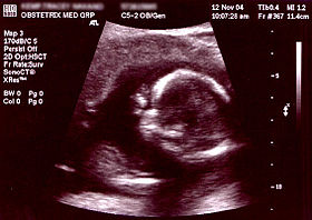
 Obstetric sonogram of a fetus at 16 weeks. The bright white circle center-right is the head, which faces to the left. Features include the forehead at 10 o'clock, the left ear toward the center at 7 o'clock and the right hand covering the eyes at 9:00. | |
| Other names | Prenatal Ultrasound |
| ICD-9-CM | 88.78 |
| MeSH | D016216 |
| OPS-301 code | 3-032, 3-05d |
Obstetric ultrasonography, or prenatal ultrasound, is the use of medical ultrasonography in pregnancy, in which sound waves are used to create real-time visual images of the developing embryo or fetus in the uterus (womb). The procedure is a standard part of prenatal care in many countries, as it can provide a variety of information about the health of the mother, the timing and progress of the pregnancy, and the health and development of the embryo or fetus.
The International Society of Ultrasound in Obstetrics and Gynecology (ISUOG) recommends that pregnant women have routine obstetric ultrasounds between 18 weeks' and 22 weeks' gestational age (the anatomy scan) in order to confirm pregnancy dating, to measure the fetus so that growth abnormalities can be recognized quickly later in pregnancy, and to assess for congenital malformations and multiple pregnancies (twins, etc).[1] Additionally, the ISUOG recommends that pregnant patients who desire genetic testing have obstetric ultrasounds between 11 weeks' and 13 weeks 6 days' gestational age in countries with resources to perform them (the nuchal scan). Performing an ultrasound at this early stage of pregnancy can more accurately confirm the timing of the pregnancy, and can also assess for multiple fetuses and major congenital abnormalities at an earlier stage.[2] Research shows that routine obstetric ultrasound before 24 weeks' gestational age can significantly reduce the risk of failing to recognize multiple gestations and can improve pregnancy dating to reduce the risk of labor induction for post-dates pregnancy. There is no difference, however, in perinatal death or poor outcomes for infants.[3]
- ^ Salomon, LJ; Alfirevic, Z; Berghella, V; Bilardo, C; Hernandez-Andrade, E; Johnsen, SL; Kalache, K; Leung, K.-Y.; Malinger, G; Munoz, H; Prefumo, F; Toi, A; Lee, W (2010). "Practice guidelines for performance of the routine mid-trimester fetal ultrasound scan". Ultrasound Obstet Gynecol. 37 (1): 116–126. doi:10.1002/uog.8831. PMID 20842655. S2CID 10676445.
- ^ Salomon, LJ; Alfirevic, Z; Bilardo, CM; Chalouhi, GE; Ghi, T; Kagan, KO; Lau, TK; Papageorghiou, AT; Raine-Fenning, NJ; Stirnemann, J; Suresh, S; Tabor, A; Timor-Tritsch, IE; Toi, A; Yeo, G (2013). "ISUOG Practice Guidelines: performance of first-trimester fetal ultrasound scan". Ultrasound Obstet Gynecol. 41 (1): 102–113. doi:10.1002/uog.12342. PMID 23280739. S2CID 13593.
- ^ Whitworth, M; Bricker, L; Mullan, C (2015). "Ultrasound for fetal assessment in early pregnancy". Cochrane Database of Systematic Reviews. 2015 (7): CD007058. doi:10.1002/14651858.CD007058.pub3. PMC 4084925. PMID 26171896.