Back نفق فرجي Arabic Canal pudend Catalan Canalis pudendalis German Conducto pudendo Spanish مجرای زهاری Persian Canal pudendal French Kanal Alcock ID Canale di Alcock Italian 음부신경관 Korean Kanał sromowy Polish
| Pudendal canal | |
|---|---|
 | |
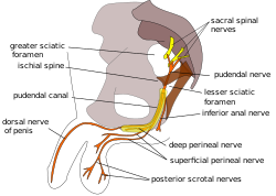
 Pudendal nerve and its course through the pudendal canal (labelled in yellow) | |
| Details | |
| Identifiers | |
| Latin | canalis pudendalis |
| TA98 | A09.5.04.003 |
| TA2 | 2436 |
| FMA | 22071 |
| Anatomical terminology | |
The pudendal canal (also called Alcock's canal) is an anatomical structure formed by the obturator fascia (fascia of the obturator internus muscle) lining the lateral wall of the ischioanal fossa. The internal pudendal artery and veins, and pudendal nerve pass through the pudendal canal, and the perineal nerve arises within it.[1]
- ^ "canalis pudendalis". TheFreeDictionary.com. Retrieved 2023-06-14.