Back Lipocalin Danish Lipocaline German Lipocalin English Lipocaline French Լիպոկաին Armenian Липокалин Russian
 | |||||||||
| Identificadores | |||||||||
|---|---|---|---|---|---|---|---|---|---|
| Símbolo | Lipocalina | ||||||||
| Pfam | PF00061 | ||||||||
| Pfam clan | CL0116 | ||||||||
| InterPro | IPR000566 | ||||||||
| PROSITE | PDOC00187 | ||||||||
| SCOPe | 1hms / SUPFAM | ||||||||
| OPM superfamily | 50 | ||||||||
| OPM protein | 1kt6 | ||||||||
| |||||||||
| Lipocalina | |||||||||
|---|---|---|---|---|---|---|---|---|---|
 Estrutura da lipocalina de Escherichia coli.[1] | |||||||||
| Identificadores | |||||||||
| Símbolo | Lipocalina_2 | ||||||||
| Pfam | PF08212 | ||||||||
| Pfam clan | CL0116 | ||||||||
| InterPro | IPR013208 | ||||||||
| |||||||||
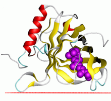
As lipocalinas constitúen unha familia de proteínas que transportan moléculas hidrófobas pequenas como os esteroides, bilinas, retinoides, así como lípidos, e a maioría das lipocalinas poden unirse ao ferro en complexos (por medio de sideróforos[2] ou flavonoides[3]) e ao hemo.[4] As lipocalinas comparten o feito de ter rexións limitadas con homoloxía de secuencias e unha arquitectura da estrutura terciaria común.[5][6][7][8][9] Esta é un barril beta antiparalelo de oito febras cunha topoloxía + 1 repetida que encerra un sitio de unión a ligando interno.[7][8]
Estas proteínas encóntranse en bacterias gramnegativas, células de vertebrados e de invertebrados e en plantas. As lipocalinas foron asociadas con moitos procesos biolóxicos, entre eles a resposta inmunitaria, o transporte de feromonas, a síntese biolóxica de prostaglandinas, a unión de retinoides e as interaccións das células cancerosas.
- ↑ Campanacci V, Nurizzo D, Spinelli S, Valencia C, Tegoni M, Cambillau C (marzo de 2004). "The crystal structure of the Escherichia coli lipocalin Blc suggests a possible role in phospholipid binding". FEBS Letters 562 (1-3): 183–188. PMID 15044022. doi:10.1016/S0014-5793(04)00199-1.
- ↑ Goetz DH, Holmes MA, Borregaard N, Bluhm ME, Raymond KN, Strong RK (novembro de 2002). "The neutrophil lipocalin NGAL is a bacteriostatic agent that interferes with siderophore-mediated iron acquisition". Molecular Cell 10 (5): 1033–1043. PMID 12453412. doi:10.1016/s1097-2765(02)00708-6.
- ↑ Roth-Walter F, Pacios LF, Bianchini R, Jensen-Jarolim E (decembro de 2017). "Linking iron-deficiency with allergy: role of molecular allergens and the microbiome". Metallomics 9 (12): 1676–1692. PMID 29120476. doi:10.1039/c7mt00241f.
- ↑ Matz JM, Drepper B, Blum TB, van Genderen E, Burrell A, Martin P, et al. (xullo de 2020). "A lipocalin mediates unidirectional heme biomineralization in malaria parasites". Proceedings of the National Academy of Sciences of the United States of America 117 (28): 16546–16556. PMC 7368307. PMID 32601225. doi:10.1073/pnas.2001153117.
- ↑ Pervaiz S, Brew K (setembro de 1987). "Homology and structure-function correlations between alpha 1-acid glycoprotein and serum retinol-binding protein and its relatives". FASEB Journal 1 (3): 209–214. PMID 3622999. doi:10.1096/fasebj.1.3.3622999.
- ↑ Igarashi M, Nagata A, Toh H, Urade Y, Hayaishi O (xuño de 1992). "Structural organization of the gene for prostaglandin D synthase in the rat brain". Proceedings of the National Academy of Sciences of the United States of America 89 (12): 5376–5380. Bibcode:1992PNAS...89.5376I. PMC 49294. PMID 1608945. doi:10.1073/pnas.89.12.5376.
- ↑ 7,0 7,1 Cowan SW, Newcomer ME, Jones TA (1990). "Crystallographic refinement of human serum retinol binding protein at 2A resolution". Proteins 8 (1): 44–61. PMID 2217163. doi:10.1002/prot.340080108.
- ↑ 8,0 8,1 Flower DR, North AC, Attwood TK (maio de 1993). "Structure and sequence relationships in the lipocalins and related proteins". Protein Science 2 (5): 753–761. PMC 2142497. PMID 7684291. doi:10.1002/pro.5560020507.
- ↑ Godovac-Zimmermann J (febreiro de 1988). "The structural motif of beta-lactoglobulin and retinol-binding protein: a basic framework for binding and transport of small hydrophobic molecules?". Trends in Biochemical Sciences 13 (2): 64–66. PMID 3238752. doi:10.1016/0968-0004(88)90031-X.