Back صبغ (علم أحياء) Arabic Tinción AST Tinció Catalan Barvení (biologie) Czech Vævsfarvning Danish Staining English Kolorigo Esperanto Tinción Spanish رنگآمیزی (زیستشناسی) Persian Coloration (microscopie) French

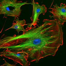
Nuclei are stained blue with DAPI, microtubles are marked green by an antibody and actin filaments are labelled red with phalloidin

A stained specimen on a glass microscope slide is mounted on the stage of a microscope
Staining is used in microscopy to make cells and tissues easier to see and understand.
This is a way to improve contrast in the microscopic image. Stains and dyes are often used in biology and medicine to highlight structures in biological tissues, often with the aid of different microscopes.
Staining can be done on living tissues (in vivo) or on dead tissues (in vitro).